The Dural Arteriovenous Fistulas (DAVF) are complex lesions involving blood vessels in the brain and less commonly the spine.
Under normal circumstances, there is gradual transition of blood flow between the arteries and veins. The arteries are designed for high blood pressure and the veins are designed for low blood pressure. This gradual transition is important because it allows for safe passage from a high pressure (arteries) to a low pressure (veins) system.
A dural arteriovenous fistula essentially means that there is abnormal connection between one of more arteries and a vein, without the gradual transition. This is called an “arteriovenous shunt”. As a result, the veins which are designed for a low-pressure system are now flooded with high pressure blood flow and show signs of dysfunction.
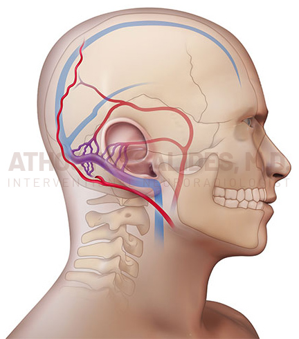
This illustration shows a dural arteriovenous fistula. The abnormal connections between arteries and veins are shown with purple color. This DAVF is in proximity to the ear and typically presents with pulsatile tinnitus.
As a result of this abnormal connection between arteries and veins (“arteriovenous shunt”), there are significant changes in blood flow involving the brain. The consequences of these changes vary significantly according to the location of the DAVF and the degree of shunt. At the good end of the spectrum, the patients may only experience pulsatile tinnitus, especially if the abnormal connection is close to the ear. At the bad end of the spectrum, the patient may eventually suffer from seizures, brain swelling (edema) or stroke.
It is therefore imperative to identify a DAVF as early as possible and consider treatment if necessary. For patients with pulsatile tinnitus, proper work-up is improtamt, as this might be the presenting symptom of a DAVF that requires treatment to prevent catastrophic consequences.
Diagnostic Work-up For FOR DAVF
A high resolution MRA with and without contrast is a non-invasive test with very high sensitivity identifying even subtle DAVF. In my practice this is the main tool to confirm or exclude DAVF.
If the MRA is suspicious for DAVF, then a minimally invasive called catheter angiogram is an important next step.
Treatment of DAVF
In the vast majority of patients, DAVF can be treated with a minimally invasive approach called embolization. The details and complexity of treatment vary depending on the location and complexity of DAVF. If embolization is not feasible or not sufficient, open surgery might be necessary.